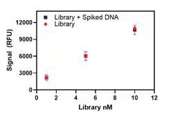

Tundra Cryo-TEM Components
Tundra Cryo TEM
Bringing cryo-electron microscopy to you
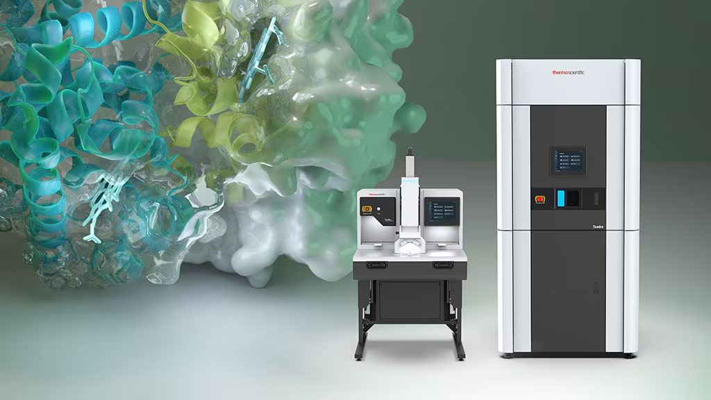
Whether you’re an experienced microscopist, or just starting out, the Thermo Scientific Tundra Cryo-Transmission Electron Microscope (Cryo-TEM) is a dedicated structural analysis solution designed to bring cryo-electron microscopy (cryo-EM) to your laboratory at an affordable price point. Simplified data collection and rapid sample loading/unloading make the Tundra Cryo-TEM a powerful tool for answering your most challenging research questions. With the Tundra Cryo-TEM, you can obtain structures at biologically relevant resolutions in-house or reliably prepare high-quality samples for further analysis on Thermo Scientific Krios or Glacios Cryo-TEMs.
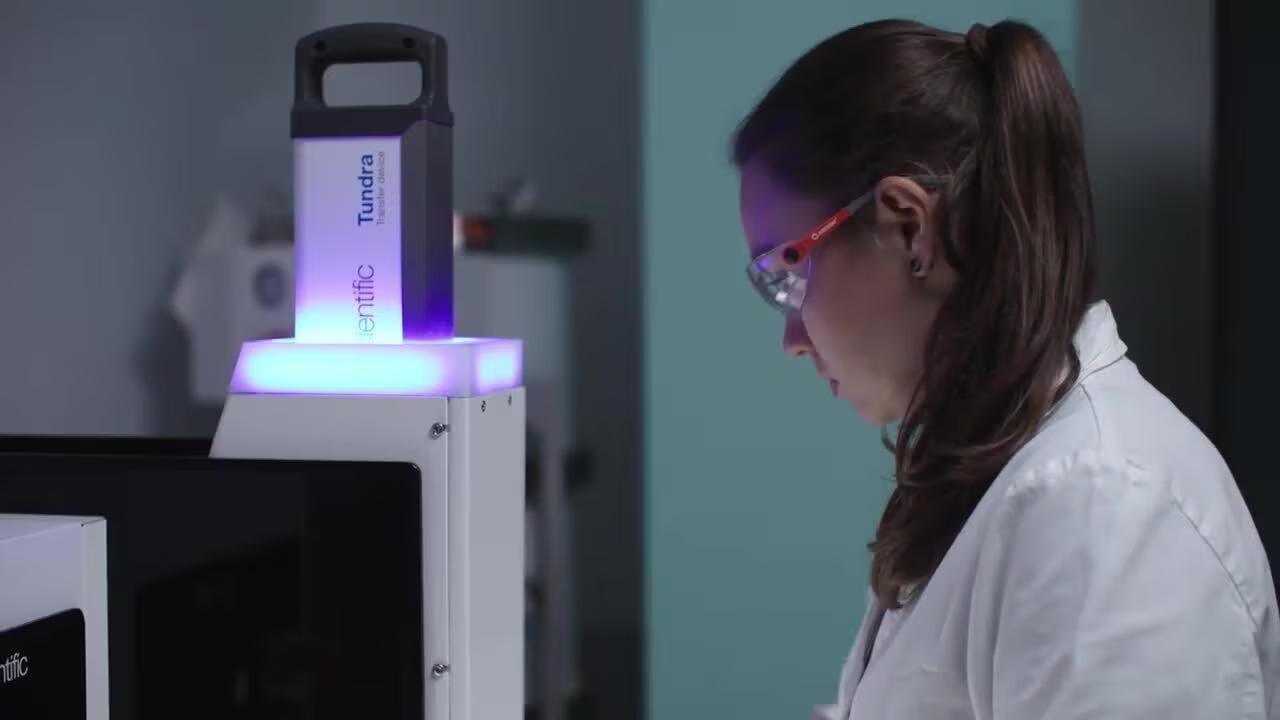
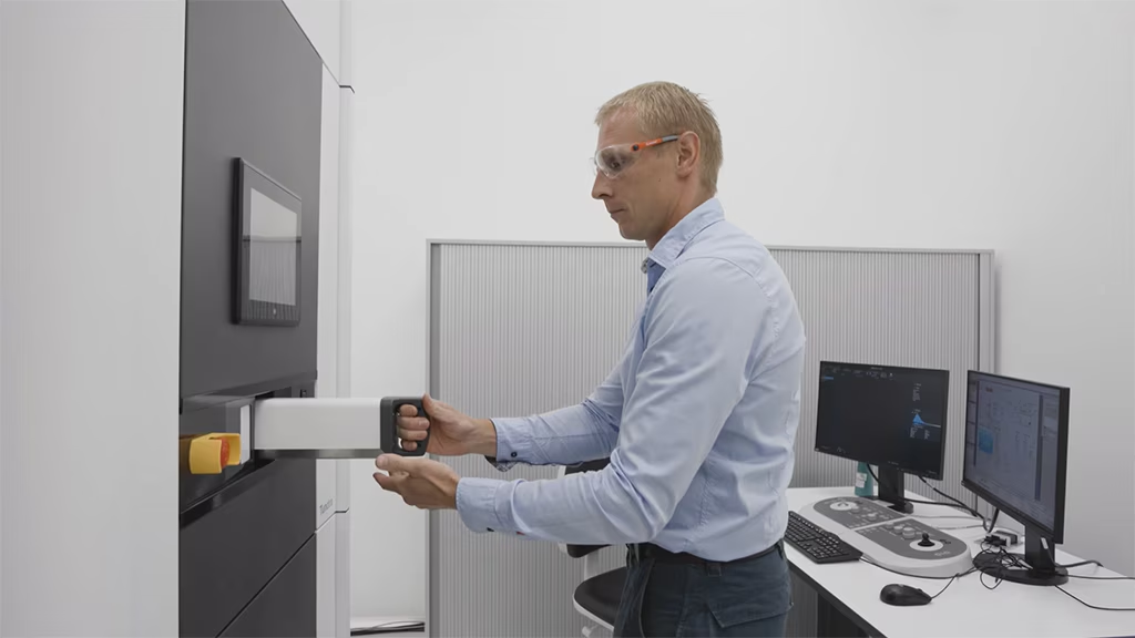
Simplified cryo-EM
The Tundra Cryo-TEM allows you to perform structural analyses on challenging proteins and macromolecules with unprecedented ease of use.
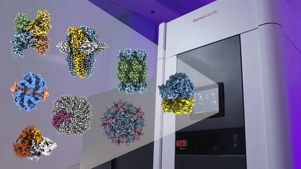
Cryo-EM structure determination at biologically relevant resolutions
The Tundra Cryo-TEM is a dedicated structure determination solution, operating at 100 kV, the Tundra Cryo-TEM can generate high-contrast biological images down to 2.1 Å.
Tundra Cryo-TEM resources
Learn more from scientists who are using the Tundra Cryo-TEM in their research and take a closer look with our on-demand product tour.
Advanced cryo-TEM software with streamlined workflows
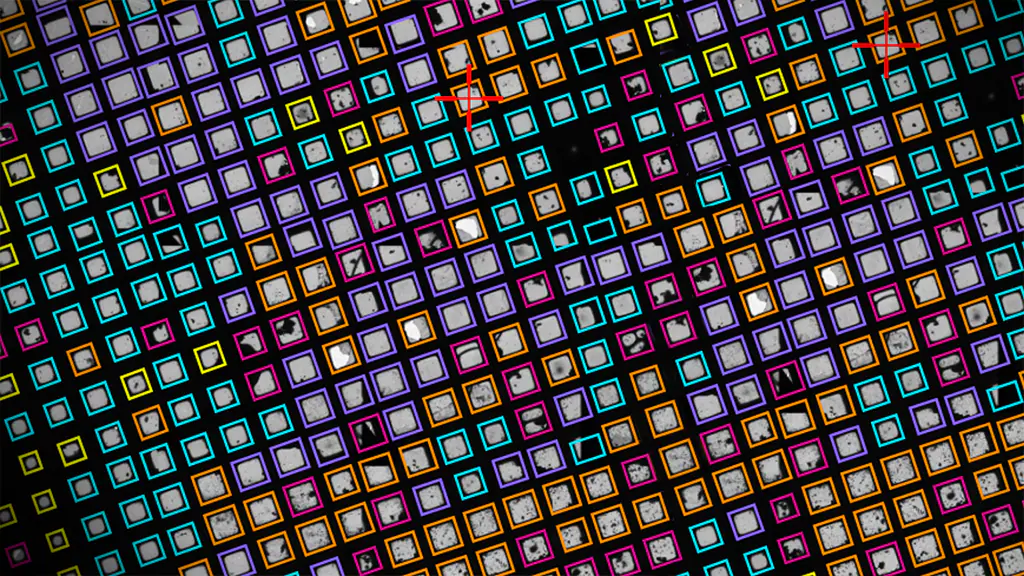
The Tunda Cryo-TEM comes with AI-enabled software to make it easier than ever to run and acquire high-resolution data.

Cryo-TEM service and support
To ensure optimal system performance, we provide you access to a world-class network of field service experts, technical support, and workflow validation.
Electron Microscopy Funding Support Center
A wide range of funding proposals and grants were considered when designing the Tundra Cryo-TEM to keep its price within the reach of most instrumentation grants.
FAQ
Frequently asked questions about the Tundra Cryo-TEM
What is the Tundra Cryo-TEM?
The Thermo Scientific Tundra Cryo-TEM is a cryo transmission electron microscope optimized for cryo-electron microscopy single particle analysis; it is designed to increase the accessibility of this powerful technique for biochemistry laboratories, pharmaceutical researchers, and more. The integrated Thermo Scientific Falcon C Direct Electron Detector allows the Tundra Cryo-TEM to perform structural analysis on a range of challenging proteins and macromolecules. With the Tundra Cryo-TEM, you can easily load and unload cryo-EM grids in a contamination-free state, discover high-resolution structures that support your biological research, and even examine room-temperature samples such as negative stain grids and plastic sections.
How does the Tundra Cryo-TEM streamline cryo-EM workflows and improve productivity?
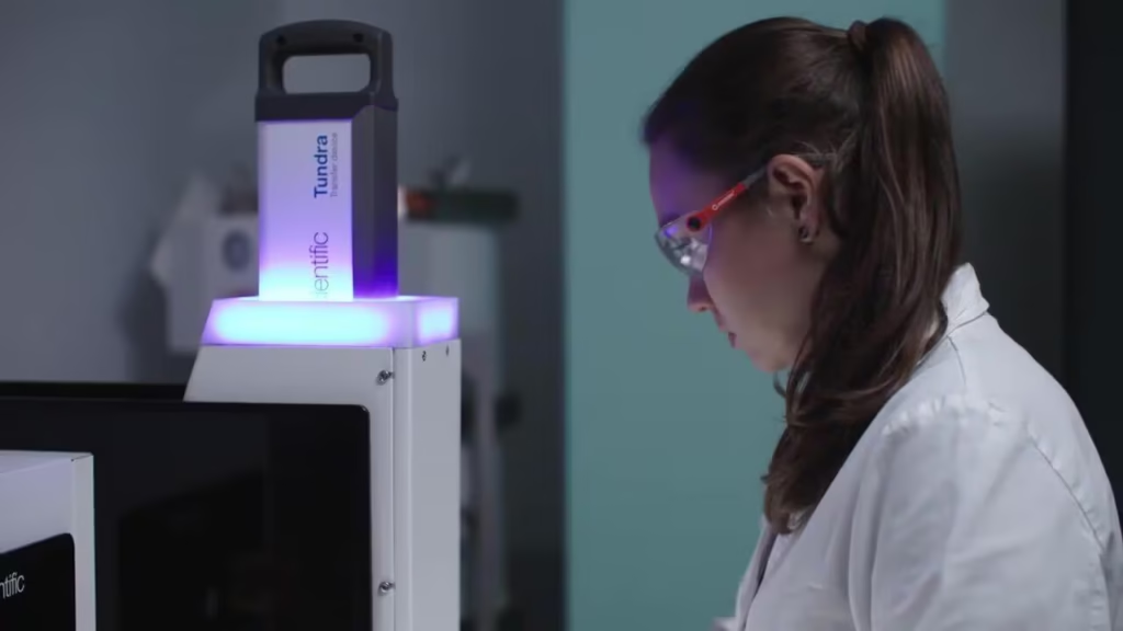
The Tundra Cryo-TEM includes a number of hardware and software features that facilitate cryo-EM workflows, thereby improving productivity. For example, it features an innovative cryo-loading station that uses automation to load the sample grid into a transfer device and then into the microscope, reducing possible user error and manual steps.
The Tundra Cryo-TEM also comes with a complete suite of automation software, including user-friendly Thermo Scientific EPU Software, which allows for high-resolution single-particle-analysis data collection in only a few clicks. The instrument is also integrated with Smart EPU Software, which is an AI-enabled solution for the analysis of intermediate results, which can be used to steer data collection on the fly.
The specialized configuration of the Tundra Cryo-TEM makes cryo-electron microscopy more accessible, facilitating the investigation of infectious and neurodegenerative diseases, cancer, and more.
How accessible is the Tundra Cryo-TEM?
The Tundra Cryo-TEM is designed with new users in mind, even those who are unfamiliar with electron microscopy. It includes a range of automation features that facilitates the entire analytical workflow, including instrument configuration, sample optimization, and data collection. Tundra Cryo-TEM data is compatible with a variety of common analytical software, supporting post-processing and reconstruction.
The Tundra Cryo-TEM only takes a few minutes to load a new cryo-sample into the microscope, allowing for rapid, automated optimization of sample conditions. This minimizes the risk of sample damage or vacuum leak from the microscope.
The rapid feedback of the Tundra Cryo-TEM significantly shortens the time required for biochemical sample optimization. This design is especially constructed so that new users are capable of doing the procedure without extensive training.
What constitutes biologically relevant cryo-EM resolutions, and does the Tundra Cryo-TEM attain these resolutions?
The Tundra Cryo-TEM can resolve protein structures down to 2.1 Å resolution through the creation of high-quality 3D reconstructions. Such high-resolution maps allow for de novo model building; the protein backbone can be clearly traced, side chains can be easily modeled, and water networks in active sites can be visualized. These biologically relevant atomic models can help you understand how proteins function, how to modify genes, and how to optimize drug designs.
Can the Tundra Cryo-TEM operate at room temperatures?
Yes, the Tundra Cryo-TEM can be operated at room temperature, supporting a number of common EM techniques. This includes negative-stain electron microscopy, which serves as an easy and cost-effective method for the assessment of purified biological specimens. Additionally, sections of resin-embedded cells and tissues can be readily visualized at room temperature.
Advanced Superparamagnetic Technology Ensures Superior Performance and Uniformity
The High-Select™ Fe-NTA Magnetic Phosphopeptide Enrichment Beads utilize cutting-edge non-aggregating, magnetite (Fe3O4) superparamagnetic technology that delivers exceptional uniformity across all applications. This advanced bead composition provides consistent and reliable performance in both manual purification protocols and automated systems, including compatibility with sophisticated instrumentation such as the Thermo Scientific™ KingFisher™ Apex Magnetic Particle Processor. The superparamagnetic properties of these magnetite-based beads ensure rapid magnetic separation while maintaining sample integrity throughout the enrichment process.
Streamlined Protocol Design Optimizes Phosphopeptide Recovery
The High-Select™ Fe-NTA Magnetic Phosphopeptide Enrichment Kits follow a systematic workflow that maximizes phosphopeptide capture efficiency. The protocol begins with standard peptide sample preparation involving lysis, reduction, alkylation, and digestion, followed by essential peptide cleanup and SpeedVac concentration steps. The magnetic beads must first be blocked to prevent non-specific binding interactions. Subsequently, peptide samples are reconstituted in binding/wash buffer and combined with the blocked beads for a 30-minute incubation period at room temperature with continuous agitation. The enrichment process concludes with magnetic collection of the beads, removal of unbound peptides, execution of multiple stringent wash steps, and final elution of the enriched phosphopeptides.
Critical Solution for Mass Spectrometry Challenges in Phosphoproteomics
Mass spectrometry (MS) represents the gold standard analytical technique for identifying protein phosphorylation sites and quantifying dynamic phosphorylation changes in biological systems. However, MS analysis of protein phosphorylation presents significant technical challenges that necessitate specialized enrichment strategies. Phosphopeptides exhibit characteristically low stoichiometry in complex protein mixtures, demonstrate high hydrophilicity that affects chromatographic behavior, suffer from poor ionization efficiency during MS analysis, and often display incomplete fragmentation patterns that complicate spectral interpretation. The inherently low relative abundance of phosphorylation modifications within complex protein samples makes enrichment an essential prerequisite for successful MS-based phosphopeptide analysis and reliable phosphoproteomics research outcomes.
features
Key features and benefits
Advanced Superparamagnetic Technology Ensures Superior Performance and Uniformity
The High-Select™ Fe-NTA Magnetic Phosphopeptide Enrichment Beads utilize cutting-edge non-aggregating, magnetite (Fe3O4) superparamagnetic technology that delivers exceptional uniformity across all applications. This advanced bead composition provides consistent and reliable performance in both manual purification protocols and automated systems, including compatibility with sophisticated instrumentation such as the Thermo Scientific™ KingFisher™ Apex Magnetic Particle Processor. The superparamagnetic properties of these magnetite-based beads ensure rapid magnetic separation while maintaining sample integrity throughout the enrichment process.
Streamlined Protocol Design Optimizes Phosphopeptide Recovery
The High-Select™ Fe-NTA Magnetic Phosphopeptide Enrichment Kits follow a systematic workflow that maximizes phosphopeptide capture efficiency. The protocol begins with standard peptide sample preparation involving lysis, reduction, alkylation, and digestion, followed by essential peptide cleanup and SpeedVac concentration steps. The magnetic beads must first be blocked to prevent non-specific binding interactions. Subsequently, peptide samples are reconstituted in binding/wash buffer and combined with the blocked beads for a 30-minute incubation period at room temperature with continuous agitation. The enrichment process concludes with magnetic collection of the beads, removal of unbound peptides, execution of multiple stringent wash steps, and final elution of the enriched phosphopeptides.
Critical Solution for Mass Spectrometry Challenges in Phosphoproteomics
Mass spectrometry (MS) represents the gold standard analytical technique for identifying protein phosphorylation sites and quantifying dynamic phosphorylation changes in biological systems. However, MS analysis of protein phosphorylation presents significant technical challenges that necessitate specialized enrichment strategies. Phosphopeptides exhibit characteristically low stoichiometry in complex protein mixtures, demonstrate high hydrophilicity that affects chromatographic behavior, suffer from poor ionization efficiency during MS analysis, and often display incomplete fragmentation patterns that complicate spectral interpretation. The inherently low relative abundance of phosphorylation modifications within complex protein samples makes enrichment an essential prerequisite for successful MS-based phosphopeptide analysis and reliable phosphoproteomics research outcomes.
Specifications
Product Specifications
Final Product Type
Peptides
Quantity
96 Reactions
Shipping Condition
Wet Ice
Workflow Step
Peptide Enrichment
Detection Method
Mass Spectrometry
Form
Solid
Format
Kit
Product Type
Fe-NTA Phosphopeptide Enrichment
Starting Material
Protease-digested Protein
Unit Size
Each
Figures
Linearity
Qubit Flex Quick Reference